How much does a PET CT scan cost?
MIC uses PET CT imaging for cardiac, oncology, and neurology applications. The Alberta Health Care Insurance Plan (AHCIP) only covers the cost of PET CT imaging in cardiac applications.
The cost of PET CT imaging in oncology or neurology imaging applications is as follows:
| PET CT Imaging Exam | Cost |
|---|---|
| FDG PET CT – Brain Scan | $999.00 |
| FDG PET CT – Standard Whole Body | $1,099.00 |
| FDG PET CT – Extended Whole Body | $1,199.00 |
| FDG PET CT – Head and Neck Package* | $1,499.00 |
| Dotatate PET CT | $3,299.00 |
| PSMA PET CT | $3,299.00 |
| Amyloid PET CT | $3,999.00 |
| Diagnostic Enhanced CT (with contrast) | $499.00 |
Please note:
- Our dual-trained radiologist and nuclear medicine physicians will protocol all requisitions to ensure an accurate price estimate.
- The price is based on the imaging application, relevant tracer, area of interest (brain, head, neck, whole body, etc.), and need for a combined diagnostic enhanced CT.
- If a combined PET and diagnostic enhanced CT is required, the CT component will be $499.
- There is no cost for PET CT imaging in cardiac applications for patients with a valid Alberta Health Care card or an out-of-province health care card (except Quebec).
- *FDG PET CT – Head and Neck Package includes a standard whole-body scan with separate, dedicated, high-resolution neck imaging.*
Patients are encouraged to talk to their employer or a Benefits Specialist before booking their exam to see if they qualify for extended coverage. Some extended benefits programs or health spending accounts may cover a portion or possibly the entire exam fee.
PET CT FAQs
Unfortunately, the rising demand for PET CT services in Canada has increased patient wait times in hospitals across the country. Depending on the indication, the waitlists for PET CT performed in Edmonton hospitals can vary. Waitlists can be in the order of weeks to months for a PET CT scan based on a triage system. Patients with more life-threatening needs will be triaged first and placed higher in the queue based on the urgency of their need for care. The waitlist for PET CT scans inside a community-based clinic like MIC is much shorter (usually a few days).
Alberta Health Care only covers the cost of PET CT imaging in a community-based clinic for heart/cardiac applications (PET CT for myocardial perfusion, viability imaging, or sarcoidosis). Unfortunately, PET CT imaging for cancer/oncology or brain/neurology applications in a community-based clinic is not covered by Alberta Health Care and must be paid for out of pocket.
Absolutely! MIC accepts all medical requisitions, and our booking team is happy to provide quotes for any PET CT exam. When speaking with our team, be sure to mention that you are travelling from outside the city/province, and we can brief you about our partnerships with two local hotels to help reduce your travel expenses (Renaissance Edmonton Airport Hotel and Delta Hotels Edmonton South).
If you intend to travel by airplane or cross the border within 24-48 hours after your PET CT scan, you should speak with a member of our team to get documentation indicating that you have had a radioactive medical procedure. Most airports and border security have sensitive radiation monitors that you may trigger if you still have minute quantities of tracer in your system. Our team can provide the proper documentation so you are not held up by security. Staying hydrated, drinking plenty of water, and voiding frequently after your appointment can also help flush the radiotracer from your body.
During a PET scan, a tiny amount of a radioactive tracer is injected into the body. The radiotracer is taken up by certain cells, where it is trapped or bound and emits small amounts of radiation, which are then detected by the PET camera. A computer uses the radiation pattern to generate an image of the tracer distribution through the body.
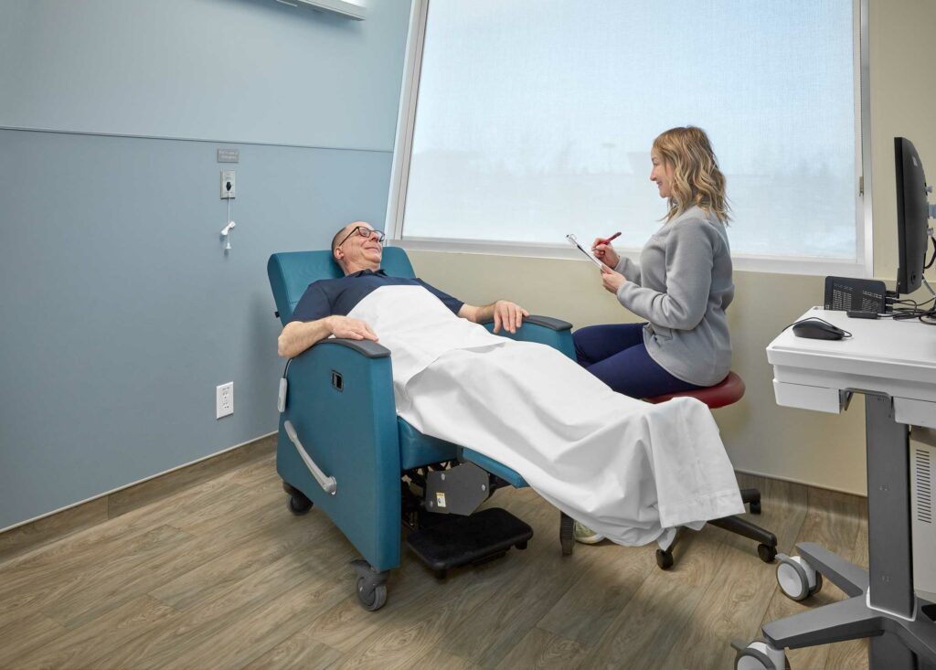
PET CT appointments consist of multiple parts, and the duration varies depending on the application or study. On average, appointments last around 2-3 hours; approximately 10-15 minutes for preparation and set up with our technologist, 30-60 minutes for the radiotracer injection/circulation, and 30-45 minutes for imaging.
A PET scan is a type of nuclear medicine imaging. PET differs from conventional nuclear medicine as the images are generated on a unique camera and often combined with other radiology studies such as computed tomography (CT).
The camera used in PET imaging can simultaneously detect two gamma-ray photons emitted from the radiotracers, whereas nuclear medicine cameras can only detect single gamma-ray photons.
The benefits of PET imaging include:
1. Lower radiation dose from smaller amounts of short-lived radiotracers.
2. Improved patient experience through a shorter exam time (minutes vs. hours or over several days).
3. Consistently high-quality images (sharper resolution with less background noise).
Our booking team will provide detailed instructions when you schedule your appointment. Unless directed otherwise, you may take medications prescribed by your physician with plain water before your exam.
In general, you should not take over-the-counter medicines like cough syrups, cough drops, or antacid tablets on the day of the exam.
If you have diabetes and fasting or an altered diet is required for your PET CT, we recommend consulting with your diabetes care provider to discuss possible medication adjustments. Typically, we instruct patients to take their insulin/oral agents at least 4 hours before the exam.
If you have any questions or concerns, please call our central booking team at 780-450-1500.
The most common concern associated with PET CT imaging is radiation exposure from the radiotracer injection.
When a PET CT scan is performed, a tiny amount of a radioactive drug, called a radiotracer, is injected into a vein. The amount of radiation from the radiotracer varies depending on the exam. For example, in cardiac perfusion imaging studies, the dose is very low and actually lower than other typical methods of imaging perfusion, such as conventional nuclear medicine.
The risk of negative effects from radiation exposure is very low. However, the tracer might:
• Expose your unborn baby to radiation if you are pregnant.
• Expose your child to radiation if you are breastfeeding.
Before we proceed with any imaging exam, your practitioner and our radiologists will review all available information to ensure the benefits from imaging will far outweigh any potential risks (such as radiation exposure).
The main side effects associated with PET CT imaging are related to intravenous contrast dye, which may be administered for the CT part of the exam. On a rare occasion, some patients may be allergic to the contrast dye.
Patients may also experience temporary side effects from the contrast dye, such as diarrhea, nausea, upset stomach, etc., which usually last less than 24 hours and subside on their own. Side effects from the radiotracer itself are extremely rare.
Having a PET CT scan while pregnant can be safe for both you and your fetus.
Generally speaking, all attempts are made to find other non-ionizing imaging methods or to delay the PET CT scan until after pregnancy. However, if the potential benefits from PET CT imaging outweigh the risks of radiation exposure, your physician may recommend the exam.
As always, your physician will help you make the best choice for your health and that of your fetus.
If you are breastfeeding, please inform your healthcare team. Occasionally, some patients may need to temporarily pause breastfeeding after their PET CT exam, depending on the type of exam and radiotracer used. Some patients may be instructed to pump before the exam or briefly use formula instead of breast milk if their feeding routines are interrupted.
When you schedule your appointment, our team will provide detailed information and discuss if any interruption is required and when breastfeeding can be safely resumed after the PET radiotracers are no longer present in your system.
When you schedule your appointment, our team will provide detailed information and discuss if any interruption is required and when breastfeeding can be safely resumed after the PET radiotracers are no longer present in your system.
PET CT imaging is essentially painless. There may be some discomfort from the injection, like donating blood. Some patients may find lying on a table for an extended period uncomfortable. Lastly, for some portions of the scan, patients may need to lift their arms above their head, which can be tiring.
No - these terms are used interchangeably by medical professionals. Nuclear medicine uses very small amounts of radioactive material, known as radiopharmaceuticals or radiotracers. Radiotracers come in different forms, such as injections, pills, and aerosol gas. In PET CT, an injectable form is used. When a radiotracer is injected into the body, it builds up in certain areas of the body that can be examined with the PET scan.
PET CT exams use a radiotracer injected intravenously before imaging. Different radiotracers have different half-lives – a half-life is the time it takes for the radioactive atoms in the tracer to decay and become non-radioactive. Some tracers will become non-radioactive faster than others and pass from the body, typically via urine or stool, within a few hours or a day.
Essentially all the radiation from a PET CT scan will be gone before the morning after your appointment. We encourage patients to drink plenty of water after their exam to help flush the radiotracer from their system. As a precaution, patients should avoid close contact with pregnant women, babies and young children for 6 hours after the scan.
Essentially all the radiation from a PET CT scan will be gone before the morning after your appointment. We encourage patients to drink plenty of water after their exam to help flush the radiotracer from their system. As a precaution, patients should avoid close contact with pregnant women, babies and young children for 6 hours after the scan.
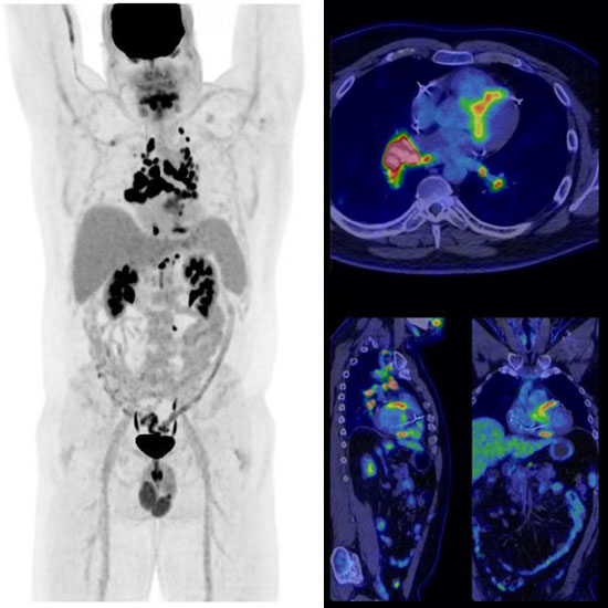
One of the most used radiotracers in PET imaging is F-18 fluorodeoxyglucose (FDG). It is just one of many radiotracer applications coming soon to MIC.
"FDG uptake" refers to the amount of radiotracer uptake that the radiologist can see on your medical images. When cells need more energy, they often use more glucose and will show more uptake on a scan. FDG uptake is particularly helpful for visualizing metabolic activity in different areas of the body.
Just because a cell is active does not mean it is abnormal. There are many reasons why cells may require more energy. For example, the brain is normally very active and shows proportionately high FDG uptake.
Sometimes there is a misconception among patients that any level of uptake is abnormal – which is not always the case and can lead to unnecessary anxiety and concern.
Physicians and radiologists are most often interested in the pattern of uptake. Some abnormal processes in the body, such as infection or cancer, cause cells to become more active, and their pattern of FDG update changes significantly.
Aggarwal, C., Henry, N., Magnuson, A., Mulrooney, D., Hlubocky, F., Patel, J., Keedy, V., Lee, R., Artz, A., Flowers, C., Markham, M., Lesser, G., Lonial, S., Pal, S., Patel, M., Rodriguez, C., Tsai, K., Yurgelun, M. (2020). Positron Emission Tomography and Computed Tomography (PET-CT) Scans. https://www.cancer.net/navigating-cancer-care/diagnosing-cancer/tests-and-procedures/positron-emission-tomography-and-computed-tomography-pet-ct-scans
Alberta Health Services. (n.d.). Positron Emission Tomography/Computed Tomography. https://www.albertahealthservices.ca/findhealth/Service.aspx?id=5931
BC Cancer. (n.d.). PET Functional Imaging. http://www.bccancer.bc.ca/our-services/services/pet-functional-imaging
Canadian Cancer Society. (n.d.). Positron Emission Tomography (PET) Scan. https://cancer.ca/en/treatments/tests-and-procedures/positron-emission-tomography-pet-scan
Cedars Sinai. (n.d.). Myocardial Perfusion PET Stress Test. https://www.cedars-sinai.org/programs/imaging-center/med-pros/cardiac-imaging/pet/myocardial-perfusion.html
Cleveland Clinic. (2022). PET Scan. https://my.clevelandclinic.org/health/diagnostics/10123-pet-scan
Columbia Radiology. (n.d.). PET-CT. https://www.columbiaradiology.org/services/pet-ct
Digirad. (2017). How is PET/CT different from traditional PET imaging? https://www.digirad.com/pet-ct-different-traditional-pet-imaging/
Health Images. (n.d.). CT Scan vs. PET Scan. https://www.healthimages.com/ct-scan-vs-pet-scan/
Johns Hopkins Medicine. (n.d.). Positron Emission Tomography (PET). https://www.hopkinsmedicine.org/health/treatment-tests-and-therapies/positron-emission-tomography-pet
Kapoor, V., McCook, M., Torok, F. (2004). An Introduction to PET-CT Imaging. https://pubs.rsna.org/doi/10.1148/rg.242025724
Mayo Clinic Medical Editors. (2023). Positron Emission Tomography Scan. https://www.mayoclinic.org/tests-procedures/pet-scan/about/pac-20385078
Memorial Sloan Kettering Cancer Center (2023). About your PET-CT with FDG Tracer. https://www.mskcc.org/cancer-care/patient-education/pet-ct-fdg
My Health Alberta. (2022). Positron Emission Tomography (PET). https://myhealth.alberta.ca/Health/Pages/conditions.aspx?hwid=aa80345
PET/CT Imaging of Berkeley. (n.d.). The Advantages of PET/CT over PET. https://petctberkeley.com/patient/petctbenefits/
Radiology Info. (n.d). PET/CT. https://www.radiologyinfo.org/en/info/pet
UCSF Department of Radiology and Biomedical Imaging. (n.d.). PET/CT Scan: How to Prepare, What to Expect & Safety Tips. https://radiology.ucsf.edu/patient-care/prepare/pet-ct