
When will I get the results from my cardiac PET CT scan?
After the cardiac PET CT scan, a dual-trained and certified MIC nuclear medicine physician and radiologist will review the images and send a detailed report to your referring practitioner, typically within one to two business days.
PET CT Cost for Cardiac Applications
There is no cost for a PET CT exam used in cardiac applications if you have a valid Alberta Health Care card (or an out-of-province health care card, except for Quebec).
Cardiac PET CT FAQs
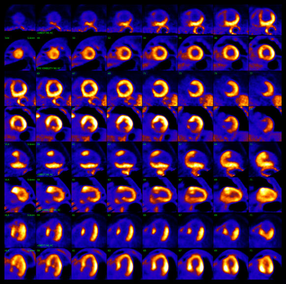
“FDG uptake” in a cardiac PET CT scan refers to the accumulation of FDG (Fluorodeoxyglucose) radiotracer within the heart muscle.
FDG is almost identical to glucose (sugar). When cells need more energy to increase metabolic activity, they often use more glucose, which is detected by medical imaging. Radiologists monitor glucose levels and tissue uptake to visualize activity in the heart.
There are many reasons why cells require more energy. Sometimes there is a misconception that any level of glucose uptake is abnormal – which is not always the case and can lead to unnecessary anxiety and concern. Your healthcare team is more interested in analyzing the pattern of uptake during a cardiac PET CT scan.
CT angiography is a non-invasive test that uses computed tomography to examine the arteries that supply blood to the heart and plaque buildup. PET MPI focuses on the blood flow to the heart during the “rest” and “stress” phases.
As MIC radiologist Dr. Abele says in an AHS press release: “In angiography, you’re basically taking pictures of the big arteries, the pipes that provide blood flow to the heart. [With PET CT] we’re imaging what actually flows out the other end. We’re measuring the actual blood flow as opposed to the size of the pipe” (2014).
As MIC radiologist Dr. Abele says in an AHS press release: “In angiography, you’re basically taking pictures of the big arteries, the pipes that provide blood flow to the heart. [With PET CT] we’re imaging what actually flows out the other end. We’re measuring the actual blood flow as opposed to the size of the pipe” (2014).
No. Patients must avoid intense or strenuous exercise such as jogging, strength training, aerobics etc. for 36 hours prior to their appointment. Excessive muscle activity can interfere with the interpretation of your images.
It depends on the specific cardiac PET CT exam. Some exams require patients to avoid caffeine outside of the fasting window. For example, patients scheduled for a PET CT for perfusion imaging must not have any caffeine for 24 hours before their exam (including drinks such as coffee, tea, energy drinks, caffeinated soda, Tylenol 3, chocolate, etc.). Our central booking team will review all the necessary exam preparation when scheduling your appointment.
As a general rule, patients should wear loose-fitting, comfortable clothing to their appointment. Depending on the exam, you may be asked to change into a gown. It is important that patients do not wear any metal during their appointment (jewelry, piercings, etc.).
MIC uses two types of radiotracers called FDG and Rubidium in cardiac PET CT applications. These radiotracers have different half-lives - a half-life is the time it takes for the radioactive atoms in the tracer to decay and become non-radioactive.
Rubidium has an extremely short half-life in the body (minutes). In under an hour, by the time your appointment is over, the tracer will be non-radioactive.
FDG also has a very short half-life in the body but will take a little longer to clear (under two hours). Essentially, by the morning after your appointment, the tracer will be gone.
Staying hydrated and drinking plenty of water after your appointment can help flush the tracer from your system.
Alberta Health Services. (2014). A quicker, safer was to ‘see’ the flow of a beating heart. https://www.albertahealthservices.ca/news/releases/2014/Page10611.aspx
American Heart Association. (2022). Positron Emission Tomography (PET). https://www.heart.org/en/health-topics/heart-attack/diagnosing-a-heart-attack/positron-emission-tomography-pet
Applied Radiology. (2009). Cardiac and neurological PET-CT applications. https://appliedradiology.com/articles/cardiac-and-neurological-pet-ct-applications
Clevland Clinic. (2023). Cardiac PET Scan. https://my.clevelandclinic.org/health/diagnostics/17376-cardiac-positron-emission-tomography-pet
Knaapen P, de Haan S, Hoekstra OS, Halbmeijer R, Appelman YE, Groothuis JG, Comans EF, Meijerink MR, Lammertsma AA, Lubberink M, Götte MJ, van Rossum AC. (2010). Cardiac PET-CT: advanced hybrid imaging for the detection of coronary artery disease. https://www.ncbi.nlm.nih.gov/pmc/articles/PMC2828569/
Memorial Sloan Kettering Cancer Center (2023). About your PET-CT with FDG Tracer. https://www.mskcc.org/cancer-care/patient-education/pet-ct-fdg
Mount Sinai. (n.d.). Cardiac PET-CT Imaging Laboratory. https://www.mountsinai.org/care/heart/services/imaging/cardiac-pet-ct
University of Ottawa Heart Institute. (n.d.). PET Myocardial Perfusion Imaging. https://www.ottawaheart.ca/test-procedure/pet-myocardial-perfusion-imaging