Medically reviewed by Dr. Eric Lenza, MDCM, MSc, FRCPC
What is nuclear medicine?
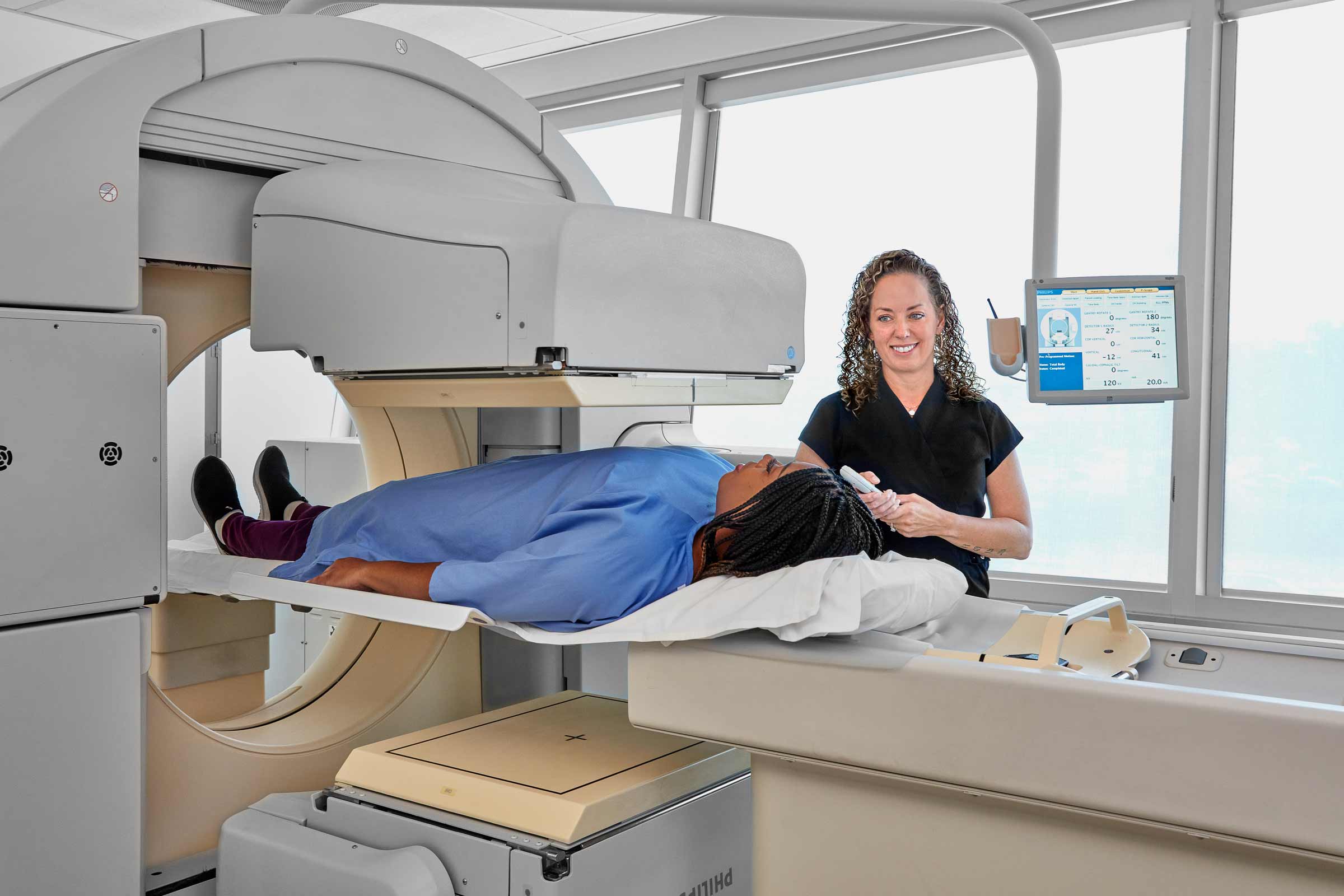
Nuclear medicine is a specialized area of medical imaging. It uses small amounts of radioactive material inside the body to create images of your organs, tissue and bones. It’s used to help diagnose and monitor a variety of health conditions.
A nuclear medicine scan shows how your organs, tissue or other body systems are working. It can help detect some diseases early, before changes are seen on other imaging tests. It’s commonly used to assess the heart, bones and kidneys, and many types of cancer. Nuclear medicine can also be used to treat certain diseases.
How does a nuclear medicine scan work?
Before your scan, you’re given a substance called a radiotracer. It contains a small amount of radioactive material. The radiotracer is typically injected into a vein in your arm. It may also be taken by mouth or inhaled, depending on the type of exam
How does the radiotracer target different parts of the body?
The radiotracer consists of two main parts. First, there’s the radioisotope that gives off radioactive energy. This radioisotope needs help to target specific areas of the body. So, it’s combined with another substance that’s naturally attracted to certain types of cells in your body. Different radiotracers are used, depending on what body part is being assessed.
How are the medical images produced?
The radiotracer travels through the body and is taken up by targeted organs or areas of the body. The absorbed tracer gives off small amounts of energy, called gamma rays. A gamma camera detects this energy, and a computer measures how much radiotracer is absorbed in different tissues. Using this information, a computer constructs detailed 3D images for our radiologists to review.
Nuclear medicine scans are often used to assess:
How is nuclear medicine different from other diagnostic imaging scans?
Function vs. Anatomy
Other diagnostic imaging scans – such as X-ray, CT, MRI and ultrasound – assess how your organs and body structures look. They evaluate your anatomy and any physical changes. Nuclear medicine shows metabolic function. Nuclear medicine scans may be combined with CT or MRI in a single exam to provide more diagnostic information.
Sources of Energy
Nuclear medicine scans also use energy in a different way. In nuclear medicine, the source of energy used to produce images comes from inside your body. That energy source is the radiotracer you’ve been given. The radiation travels out of your body and into the scanner.
X-ray, CT, MRI and ultrasound, on the other hand, use an external energy source to form pictures. This energy travels from the machine and into your body. In X-ray and CT, small amounts of radiation are directed into your body. Ultrasound uses sound waves to generate images. MRI combines a strong magnetic field with radio waves to produce pictures.
Is there a “best” type of imaging?
One type of diagnostic imaging is not necessarily better than another. Each has advantages and limitations. Your healthcare practitioner will decide which type of scan you need, depending on the body part and health condition being assessed. You may also be sent for more than one test if more diagnostic information is needed.
How safe is the radiation?
The amount of radiation you receive is low, although more than a typical X-ray. Radiotracers are carefully selected to obtain optimal imaging while minimizing radiation exposure. When sending you for imaging, your healthcare practitioner will weigh the benefits vs. risks.