Lung VQ Exam Prep
Find out when you book
Lung VQ Locations
Find out when you book
Medically reviewed by Dr. Eric Lenza, MDCM, MSc, FRCPC
What is a lung VQ scan?
A lung VQ scan is a nuclear medicine test of the lungs that checks airflow (ventilation) and blood flow (perfusion). A Lung VQ scan is most often used to diagnose a blood clot in the lungs, or pulmonary embolism.
How does a lung VQ scan work?
A lung VQ scan uses a small amount of radioactive material to produce images. As the radiotracer passes through your lungs, it gives off radiation called gamma rays. Gamma cameras pick up the signals, and a computer generates images
Comparing the Images
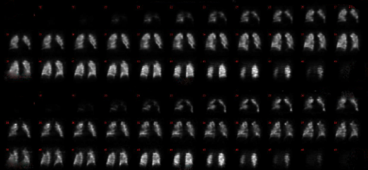
Our radiologist will compare the two sets of images. Healthy lungs should have roughly the same amount of air and blood flow. If there’s a difference or mismatch, you may have a blood clot.
How do I prepare for my lung VQ scan?
No preparation is necessary. You may eat and drink as normal before your scan.
What to Expect During a Lung VQ Scan
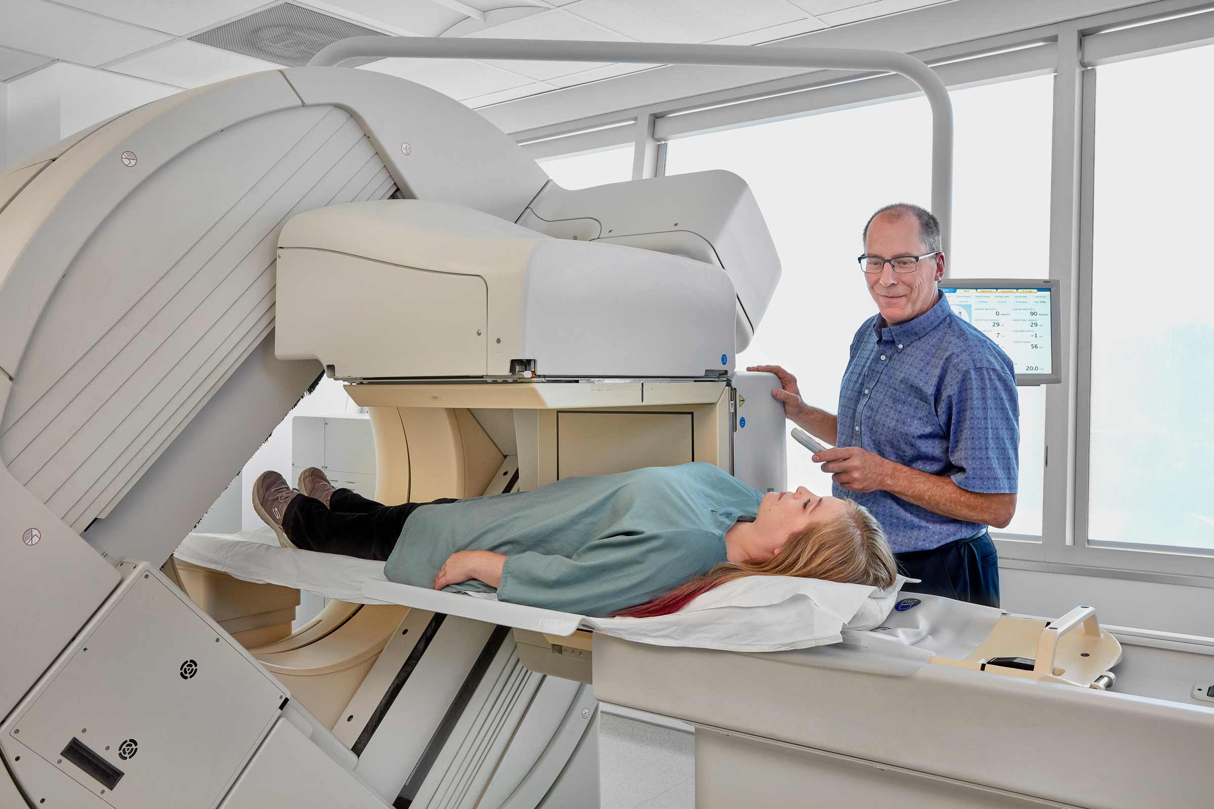
A lung VQ scan is done in two steps, back-to-back. For both parts, you will lie down on the scanning bed while the cameras take pictures. The entire exam takes about 45 minutes.
Step One: Ventilation
- The first part checks how air is moving through your lungs.
- The radiotracer comes in gas form.
- You’ll hold a tube to your mouth and breathe in the gas.
- As the radioactive material passes through your lungs, the cameras take pictures to show where air is flowing.
Step Two: Perfusion
- The second part checks blood flow through the tiny vessels, or capillaries, in your lungs.
- This time, a radiotracer is injected into your arm.
- It contains tiny particles of radioactive material. They’re small enough to accumulate in your lung capillaries.
- The cameras will take another set of pictures.