Arthrogram Exam Prep
Find out when you book
Arthrogram Locations
Find out when you book
Medically reviewed by Dr. Eric Lenza, MDCM, MSc, FRCPC
What is a nuclear medicine arthrogram?
An arthrogram is an imaging test of your joints. It can be used to assess different areas of your body, such as hips, knees, shoulders and elbows. Depending on what is being assessed, arthrograms can be performed using nuclear medicine, CT, or MRI.
A nuclear medicine arthrogram is mainly used to evaluate prosthetic joints, such as hip replacements, to assess implant loosening.
How does an arthrogram work?
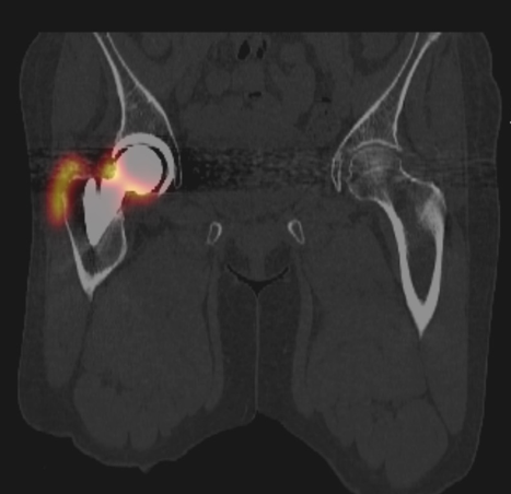
A small amount of radioactive material is injected into the joint space. The cameras detect the radiation, and a computer uses the information to generate images. If radioactive material is seen around the joint implant, it means the implant is loose.
How do I prepare for an arthrogram?
No special preparation is required. The entire procedure takes about 2 hours.
What to Expect During an Arthrogram
- You may be asked to change into a gown when you arrive for your exam.
- First, the radioactive tracer is injected directly into your joint space. This is done under the guidance of a fluoroscopy machine. Fluoroscopy is a real-time X-ray that allows our radiologist to see exactly where the needle is going.
- You’ll be asked to walk around for 30 minutes. This gives the radiotracer time to flow through the joint space.
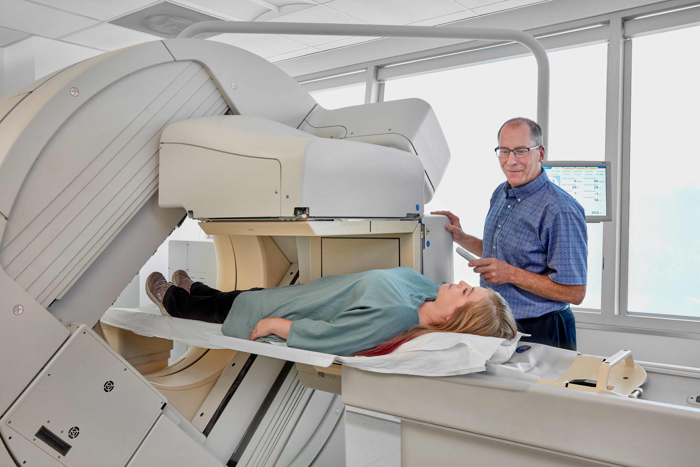
- Then, you’ll lie down on the nuclear medicine scanning bed.
- The cameras will take a series of images over a 1-hour period.
- The entire procedure takes about 2 hours.