For more patient information on Digital Breast Tomosynthesis
Mammography Exam Prep
Find out when you book
Breast Imaging Locations
Find out when you book
What is mammography?
Mammography is a specific type of imaging that uses a low-dose x-ray to examine breast tissue. A mammography exam, called a mammogram, is used to aid in the early detection and diagnosis of breast diseases.
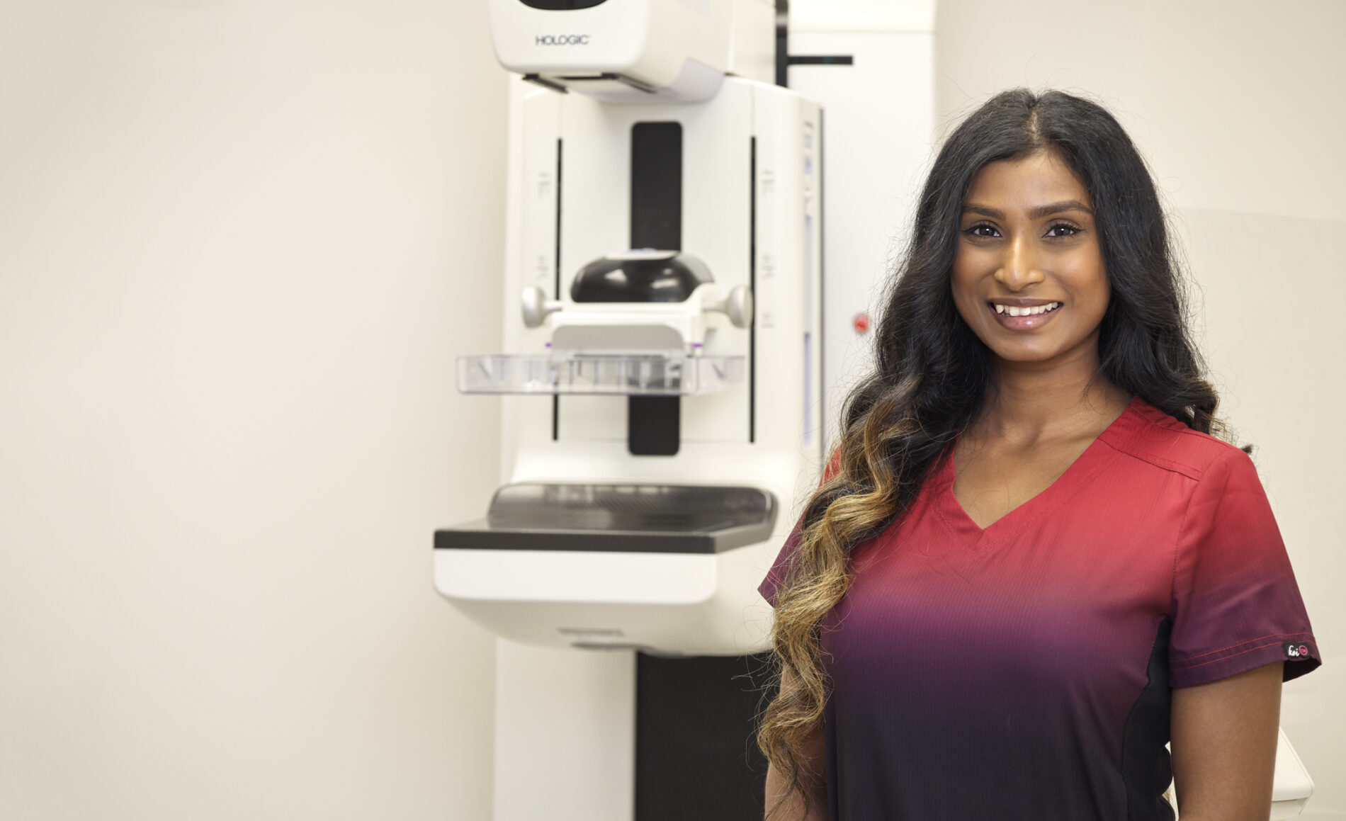
Digital Breast Tomosynthesis (DBT) Mammography
MIC uses Digital Breast Tomosynthesis (DBT) for all mammograms. DBT is an advanced form of mammography that combines a low-dose x-ray system and computer reconstructions to create three-dimensional images of the breasts.
DBT has been shown to improve cancer detection rates while reducing the number of patients recalled for additional imaging and biopsies.
Screening Mammography
Mammograms are used as a screening tool for early detection of breast cancer before any symptoms. Screening mammography can detect cancer up to two years before any breast changes are noticeable to a patient or healthcare practitioner.
Diagnostic Mammography
Diagnostic mammography uses the same Digital Breast Tomosynthesis technology as screening mammography. It’s called a diagnostic mammogram when it’s used to evaluate a patient experiencing symptoms. These symptoms can include a breast lump, pain, nipple discharge, or other changes. Diagnostic mammography may also be done after an abnormal screening mammography to further evaluate the area of concern. It focuses on a specific area of the breast and can provide more detailed images.
What to expect
- When you arrive for your appointment, you will be asked to change into a gown.
- To achieve the clearest picture, the technologist will position your breast on a compression plate and the machine will gradually compress your breast between two x-ray plates. This may cause you some temporary discomfort but it is important to flatten out the breast to get the clearest picture of the breast tissue using the least amount of radiation.
- This procedure takes only a few seconds for each image, and any tightness or discomfort you may feel usually disappears immediately after the compression is released.
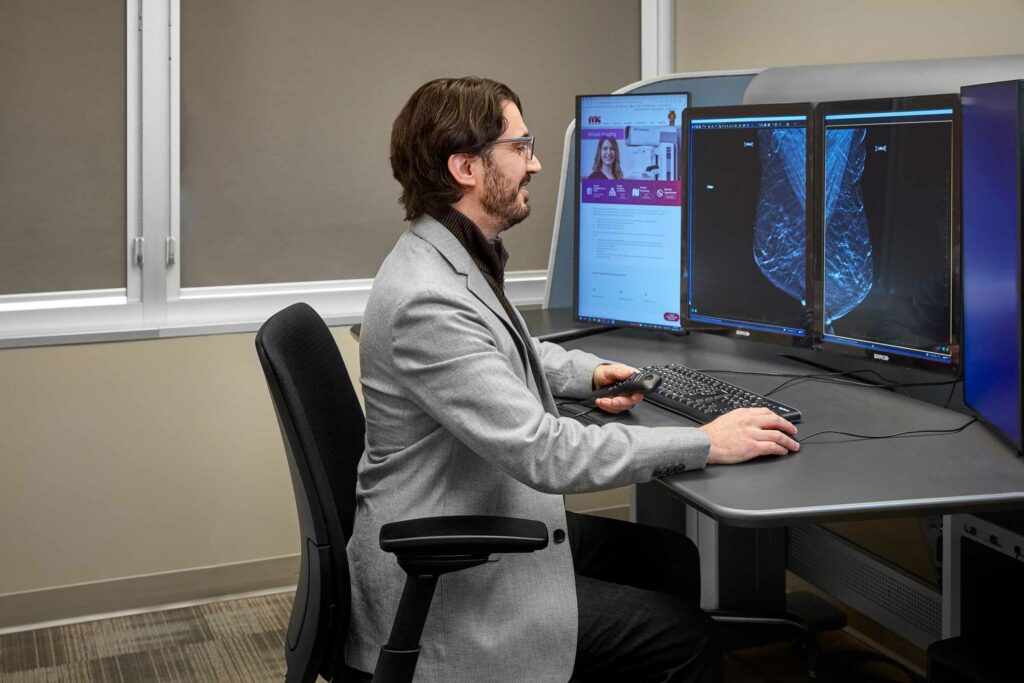
- After the radiologist has a chance to thoroughly study your images, a complete report will be sent to your referring practitioner, usually within 24 hours.
*Diabetic patients who use a glucose monitoring device should check with their manufacturer to see if they recommend removing their device before low-dose radiation exposure.