Our Central booking line will be closed on Saturday, September 19 to accommodate a staff learning and education session. You can manage your bookings online using Patient Connection, or call when regular hours resume on Monday September 21.
Myocardial Perfusion Imaging (MPI), also known as a MIBI, is a type of nuclear medicine test that checks blood flow to your heart. It can show how well different areas of your heart are working.
An MPI exam may be ordered to:
Diagnose the cause of chest pain or pressure
Check for coronary artery disease
Assess damage to heart muscle after a heart attack
Evaluate blood flow after a heart procedure, such as a stent or bypass
How does an MPI exam work?
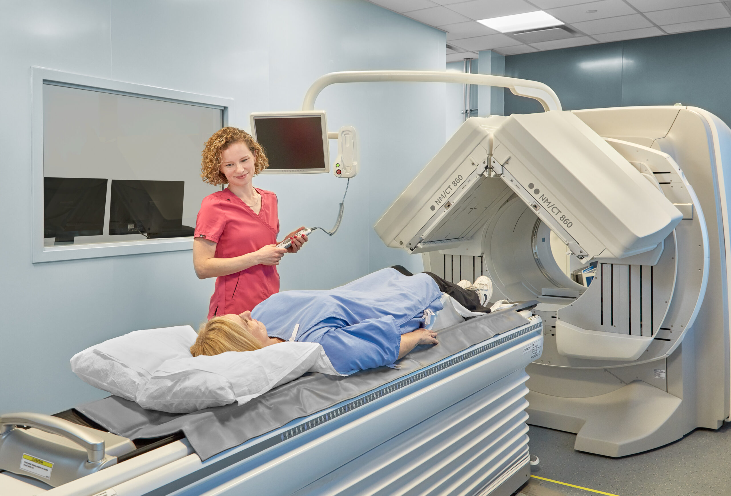
An MPI exam uses a low dose of radioactive material to produce 3-D images. The radiotracer is injected into your arm and circulates through your heart. As it passes through the arteries in your heart, it’s absorbed by healthy surrounding tissue.
How does it assess my heart?
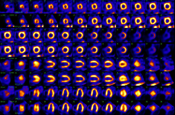
The radiotracer gives off radiation called gamma rays. Gamma cameras detect those signals, and a computer uses the information to produce images of your heart. Those images show blood flow into different parts of the heart muscle. In areas with reduced or no blood flow, the radiotracer will not appear. Those areas will appear as dark spots on the image.
How should I prepare for my MPI test?
When you book your appointment, our Central Booking team will provide instructions on diet and medications. It is important that you follow these closely. The test will take place over two days to allow MIC to image your heart after exercising and again while resting. Each appointment takes approximately 2-3 hours.
Diabetic patients who use a glucose monitoring device should check with the manufacturer to see whether they recommend removing the device before low-dose radiation exposure.
Day One: “Stress Test”
Nothing to eat or drink, except water, for four hours before
No caffeine for 24 hours prior to exam (including chocolate, cola, decaffeinated beverages, teas, etc.)
Wear clothing suitable for exercise (e.g. jogging pants or shorts, and runners)
Some medications may not be taken prior to the exam. Bring a high-fat snack to eat. The fat content can help with image quality. There may be a wait between the stress test and imaging.
Detailed instructions will be given at the time of booking
Day Two: “Rest Test”
Nothing to eat or drink, except water, for four hours before
What to Expect During an MPI Exam
Myocardial perfusion imaging is a two-part exam. You will have to come to the clinic on two different days.
Day One: “Stress Test”
The first day is the “stress test.” Your heart will be imaged after it has been stressed – either by treadmill exercise or by medication. During the exam, you’ll be monitored by a cardiologist and a technologist.
This appointment will take between 30 minutes to 1.5 hours.
Our staff will ask questions about your medical history and explain the procedure.
Before getting on the treadmill, you’ll require some preparation. Our technologist will attach electrocardiogram wires to your body with stickers.
An intravenous line will be inserted into one arm. This is how the radiotracer will be injected during the test.
A blood pressure cuff will go around your other arm.
Next, you’ll start walking on the treadmill. Every three minutes, the speed and incline will increase.
You’ll be asked to keep moving until you reach a target heart rate. Then you’ll be taken to the scanner, which will capture images of your heart.
If you are unable to exercise on a treadmill, you may be given a medication that dilates the blood vessels in your heart. This produces a similar effect to exercise.
Day Two: “Rest Test”
The second day is the “rest test.” Your heart will be imaged while you are at rest. Our radiologists will compare the images to assess your heart health.
This appointment will take between 45 minutes to 1.5 hours.
On the second day of the exam, you’ll be given another injection of the radiotracer.
The scanner will take pictures of your heart at rest.
After your test, you can go back to normal activities right away.
Drink plenty of water to flush the radioactive material from your body.
Our radiologist will review all of the information and send your practitioner a complete report, usually within 24 hours.
Why are “stress” and “rest” images needed?
When you’re relaxed, your body needs less oxygen. Your body gets oxygen from blood pumped by your heart. Since your heart is not working as hard when you’re not moving, the heart muscle also needs less blood. Blood flow may still be enough, even if the arteries in your heart are partially blocked.
When you’re exercising, your body requires more oxygen. Your heart has to work harder to pump oxygenated blood to the rest of your body. If your arteries are partially blocked, your heart muscle won’t get the blood it needs. On your MPI scan, the radiotracer won’t show up in those areas.
If parts of the heart muscle have died, those areas will not light up – during stress or rest.
Our radiologist will compare your “stress” and rest images to assess blood flow to your heart. The test also shows how well your heart is pumping.
What to expect after my MPI?
All of the radiation in your body should be gone within 24 hours. Most of the radioactivity disappears on its own. The rest is cleared from your body through urine.